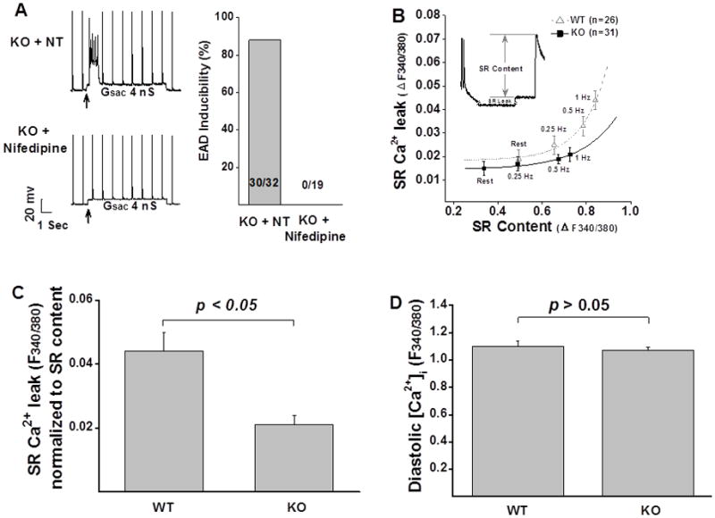
Figure 2. Cellular substrates that affect abnormal impulse induction.

A: Low concentration of the LTCC blocker nifedipine eliminated abnormal impulses in KO myocytes. B: SR Ca2+ leak was reduced in KO myocytes at all tested frequencies, compared to that recorded from WT myocytes. C: Mean values of normalized SR Ca2+ leak measured in WT (n=10) and KO (n=15) myocytes. D: There is no difference in diastolic Ca2+ between WT (n=18) and KO myocytes (n=24). Error bars denote S.E.M.; NT: normal Tyrode’s solution.
