
Figure 4. Structure of the Fbxl3 LRR domain.
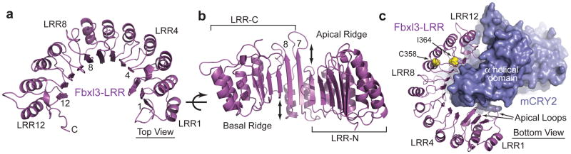
a, Ribbon diagrams of the LRR domain of Fbxl3 along with its complete C-terminal tail. Select LRRs are labeled and numbered at their helices and β-strands. b, An orthogonal view of the LRR domain shown in Fig. 4a. Double-ended arrows indicate the offset between LRR6 and LRR7 and between LRR8 and LRR9. c, A bottom view of the Fbxl3-LRR domain (magenta) bound to mCRY2 (blue). Side chains of the Fbxl3 residues mutated in the overtime and after hours alleles are shown in yellow spheres.