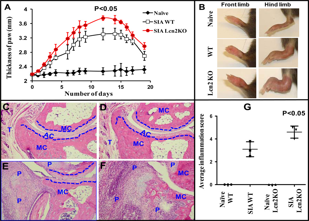
Figure 5. Lcn2 deficient mice develop severe arthritis when compared to WT in SIA model.
SIA was induced as described under Materials and Methods. The mice were monitored for the development of arthritis by ankle thickness (A) and photographs were taken on day 12 (B). Histological section of ankle joints stained with haematoxylin and eosin (original magnification 10x). Tissue sections of hind limbs from naïve WT (C) and Lcn2KO mice (D) show remarkably intact bone structure. Tissue sections of arthritic hind limbs exhibit greater infiltration of immune cells, bone erosion and cartilage destruction in Lcn2KO (F) compared to WT mice (E). Histological scoring and assessment of bone erosion in arthritic paws of Lcn2KO and WT mice were carried out as described under Materials and Methods and represented graphically (G). The blue dotted line differentiates the metacarpal (MC) bone from articular cartilage (AR). P and T stand for panus formation and tendon muscle, respectively. A group of mice (n=3) treated with PBS (naïve) served as specificity control. Photographs are representative of three individual mice. Data are expressed as ± SEM.

