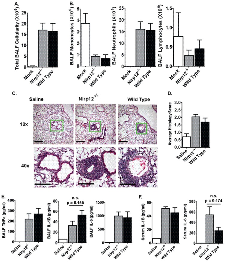
Figure 2. LPS mediated acute airway inflammation in Nlrp12−/− mice.
Mice were challenged with 1 mg/kg of E. coli LPS (serotype 0111:B4) i.t. and airway inflammation was assessed 48 hours post-challenge. A) LPS induced a significant increase in BALF cellularity. B) No significant differences were detected in the cellular composition of the BALF between Nlrp12−/− and wild type mice. C) Airway challenge with LPS resulted in a significant influx of neutrophils to the airway. Histopathology revealed a significant amount of perivascular and peribroncholar cuffing and some slight alveolar occlusion following lung LPS exposure. The magnification bar at 10× = 10 µM and 40× = 5 µM. D) Histopathology analysis and histology scoring revealed no significant differences between the Nlrp12−/− and wild type mice. E–F) LPS induced a significant increase in local (BALF) and systemic (serum) levels of proinflammatory cytokines. No significant differences were observed between Nlrp12−/− and wild type mice. Saline, n = 3; LPS-treated Nlrp12−/−, n = 6; LPS-treated Wild Type, n = 6. Experiments were repeated 3 separate times with the same numbers of mice in each replicate.

