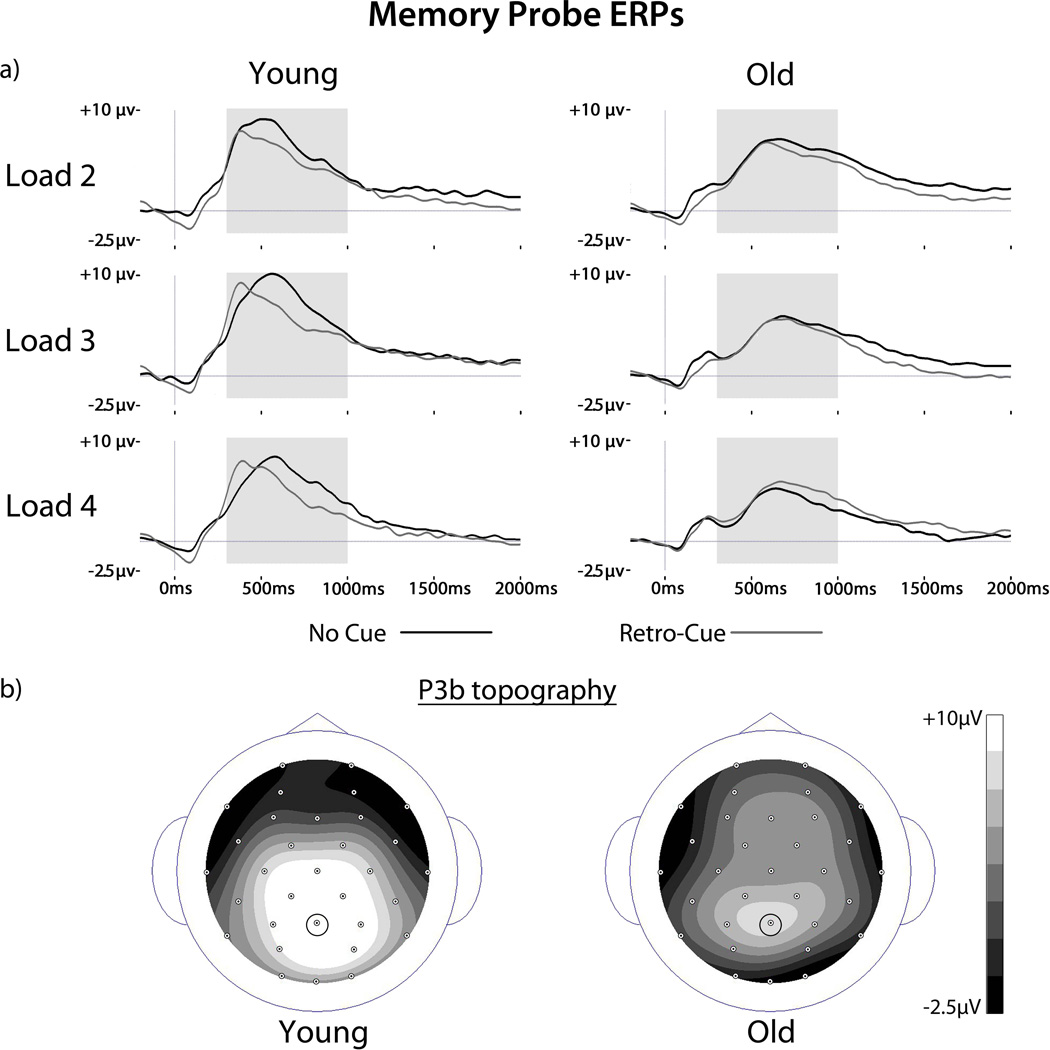
Figure 3.
ERPs to memory probes. (A) ERPs are shown for no cue and retro-cue targets for each set size and age group at electrode Pz. The P3b component is clearly visible for all conditions and age groups beginning at ~300 ms post probe onset. Gray shading indicates the time windows for P3b analysis. (B) Scalp topographies of the P3b collapsed across load and cue type are shown for each age group. Small circles represent electrode locations as viewed from above. The Pz electrode chosen for P3b analysis is indicated.

