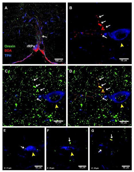
Figure 9. DMH-orexin neurons project to serotonin neurons in the rRPa in lactating rats.
(A) 20x confocal image showing triple label immunostaining for orexin (green), BDA (red), and TPH (blue) in the rRPa. The arrow indicates a fiber containing orexin and BDA near a TPH-expressing neuron. (B) 40x confocal image showing BDA-labeled axonal swellings (white arrows) in the vicinity of a TPH neuron (yellow arrowhead). (C) 40x confocal image showing orexin immunoreactivity in the same section. (D) Overlay image indicating colocalization of BDA and orexin immunoreactivities in the same axonal swellings in the vicinity of this TPH neuron. (E-G) Examples of single, confocal optical slices (1 μm thickness) illustrate colocalization of BDA and orexin immunoreactivities in axonal swellings that are in close apposition to a TPH-expressing neuron.

