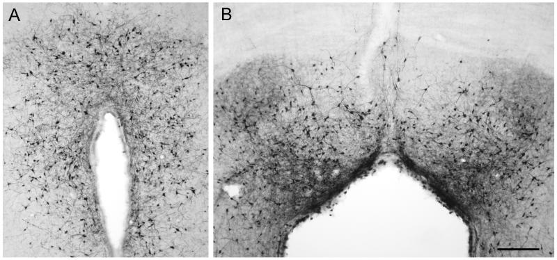
Figure 1. AAV2.5 can cross the ependymal cell barrier and transduce neurons after ventricular administration.
AAV2.5/GFP (10 uL, 6.6×10¹⁰ vg) was injected into the anterior portion of the right lateral ventricle of adult rats, and after 2 weeks gene expression was assessed by anti-GFP IHC. (A) Transduction of cells with neuronal morphology in the hypothalamus along the third ventricle. (B) GFP-positive cells with neuronal morphology in the dorsal central gray. Scale bar is 50 microns.

