
Figure 4. The NHP spinal cord and DRG are efficiently transduced following intrathecal AAV9 vector administration.
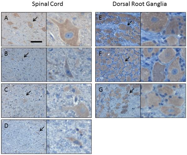
Four weeks following intrathecal injection of scAAV9/CBh-GFP vector, 5-7 micron sections from the spinal cord and DRG were subjected to IHC against GFP. Shown are sample images from those used for histomorphetric quantitation in Figure 3, and more information is available in Supplemental Figure 6 and Supplemental Table 2. These images are from NHP 203 and the specific areas are as follows, with spinal cord in panels A-D and DRG in panels E-G: (A) cervical, ventral gray matter, (B) thoracic, ventral white matter, (C) lumbar, dorsal gray matter, (D) lumbar, dorsal white matter, (E) cervical, (F) thoracic, and (G) lumbar. For each panel, the right image is a 4x enlargement of the area indicated by an arrow, and the total width of the right field is 55 microns. Slides are counterstained with cresyl violet (blue) to show nuclei, and brown (DAB) indicates GFP expression. Scale bar in (A) is the same for all panels and is 55 microns.