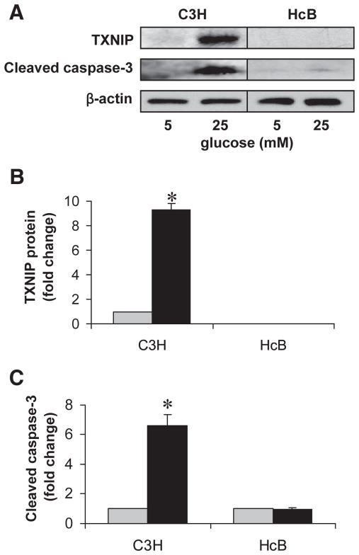
FIG. 3.
Effects of high glucose exposure on caspase-3 activation in TXNIP-deficient HcB-19 and C3H control islets. Isolated primary islets of control C3H and TXNIP-deficient HcB-19 mice were incubated at low (5 mmol/l) or high (25 mmol/l) glucose for 24 h and assessed for TXNIP expression and apoptosis. A: Representative immunoblot. B: Quantification of TXNIP protein levels in C3H and HcB-19 islets. (As expected, no TXNIP protein was detected in HcB-19 islets.) *P < 0.005. C: Quantification of cleaved caspase-3 in C3H and HcB-19 islets. Bars represent mean fold change ± SE in protein levels corrected for β-actin (n = 3 independent experiments). *P < 0.05 high vs. low glucose.
 , 5 mmol/l glucose; ■, 25 mmol/l glucose.

