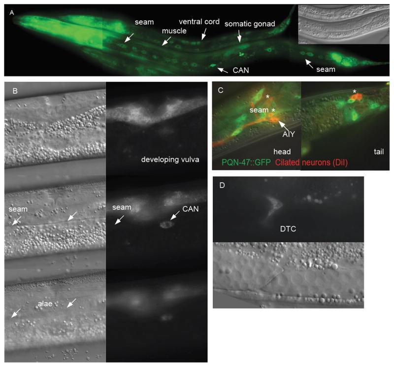
Figure 4. PQN-47∷GFP is expressed in many tissues.

(A) L1 and L2 animals in a long exposure image to show PQN-47∷GFP expression (translational fusion) outside the pharynx and tail. Inset Nomarski image shows gonad for staging (B) 3 serial focal planes showing that once seam cells have made alae (left lower NOM image), they no longer express PQN-47∷GFP (see focal plane matched GFP image on lower right). Middle left nomarski (NOM) shows seam cells in focus, though no GFP expression (focal plane matched GFP). PQN-47∷GFP is however expressed in developing (L4) vulval cells (top row of images). (C) PQN-47∷GFP is expressed in senosry neurons in head and tail. The head cells were identified by position as chemoattractive ASI, AKS and/or chemorepulsive neuron ADL or sheath cell AWB and the tail neurons are chemorepulsive neurons PHA and PHB. (D) migrating distal tip cells.
