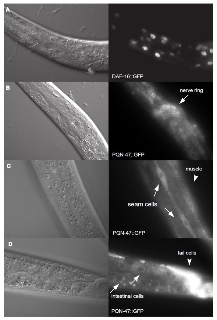
Figure 6. Leptomycin B, an inhibitor of export from the nucleus, does not drive PQN-47∷GFP into the nucleus.

Nomarski images taken at the same focal plane as the florescent images to the right. (A) After 24 hours of LMB treatment 20 out of 20 worms observed had DAF-16∷GFP in the nucleus (representative image is shown), whereas DAF-16∷GFP was cytoplasmic in 20 out of 20 animals treated with methanol control (counted on a dissecting scope). DAF-16∷GFP begins to accumulate in the nucleus upon mounting on a slide and imaging, so over time the even the methanol control treated worms turns positive, as the stress of paralysis and imaging drives it into the nucleus (no image shown). Three hours of LMP treatment is sufficient to drive DAF-16∷GFP into the nucleus of hyp7 and intestinal cells in 9 out of 10 worms counted. Synchronized L1s are small but superficially healthy worms after 24 hours on LMP treatment compared to the methanol controls (personal observation). (B) PQN-47∷GFP was not seen in the nucleus at 24 hours of LMB treatment, nor under any conditions or cells examined. (C) Even in the larger seam cells, there is little PQN-47∷GFP in the nucleus, even after 3 hrs of LMB treatment, when 12 out of 13 animals have entirely nuclear localization of DAF-16∷GFP. (D) Although there is only faint cytoplasmic PQN-47 expression in the intestine, it remains cytosolic after 3 hrs of LMB treatment.
