
Fig. 5.
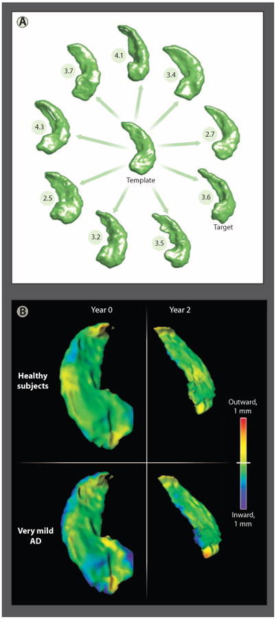
Methods of computational anatomy. (A) A sample of hippocampi from a population of healthy subjects (n = 57) and subjects with very mild AD (n = 38) (88). The anatomical template was generated from all hippocampi from this study. Also shown are the distances between nine individual hippocampi selected from the population and the template. (B) Patterns of hippocampal shape change over a 2-year period in healthy elderly subjects and subjects with very mild AD (90). The shape and volume changes revealed using CA methods support the detection of AD onset.