FIG. 2.
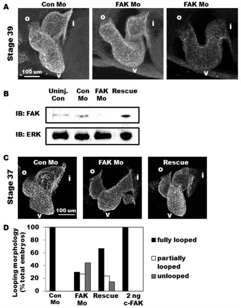
FAK morphant embryos exhibit marked cardiac dysmorphogenesis. A: Lateral view of whole-mount immunohistochemistry for tropomyosin reveals a fully looped three-chambered heart in Con Mo-injected embryos while those injected with FAK Mo appear distended and partially looped (middle panel) or unlooped (right panel). Anterior is to the left and dorsal toward the top in all panels. Regions of interest are labeled as follows: i, inflow tract; v, ventricle; o, outflow tract. B: Western blot analysis for FAK in uninjected, Con Mo-injected, FAK Mo-injected, and rescue embryos at Stage 37. Levels of ERK are shown as a control for loading. C: Lateral view of whole-mount immunohisto-chemistry for MHC reveals rescue of the FAK morphant phenotype is achieved by coexpression of 2 ng chicken FAK. D: Heart morphology analysis of Con Mo-injected, FAK Mo-injected, rescue (FAK Mo and 2 ng chicken FAK co-injection), and 2 ng chicken FAK alone demonstrates that while FAK morphant embryos exhibit full looping in only 30% of embryos examined, rescue embryos exhibit full looping morphology in 67%. Total number of embryos analyzed were n = 27 (Con Mo), n = 34 (FAK Mo), n = 42 (Rescue), n = 20 (2 ng c-FAK), collected from two separate experiments.

