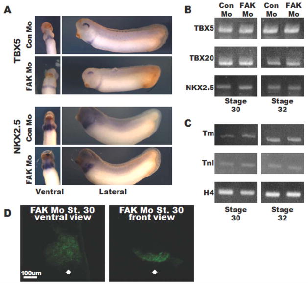
FIG. 3.
FAK depletion does not impact cardiac specification or differentiation. A: In situ hybridization of Stage 30 embryos for TBX5 and NKX2.5. Ventral views (left panels) are oriented with anterior toward the top, lateral views (right panels) are oriented with anterior to the left and dorsal to the top. B, C: RT-PCR analysis at Stages 30 (left panels) and 32 (right panels) for TBX5, TBX20, NKX2.5 (B), and tropomyosin (tm), and Troponin T (TnT) (C). Histone H4 (H4) serves as a control. Data represent results from 10 embryos per condition and experiments were repeated at least twice. D: Ventral and front views of whole-mount immunohistochemistry for MHC reveals a continuous and fused sheet of differentiated myocytes at the ventral midline (indicated by white arrow) in FAK Mo-injected embryos. Images represent 3D reconstructions of confocal z-stack sections.

