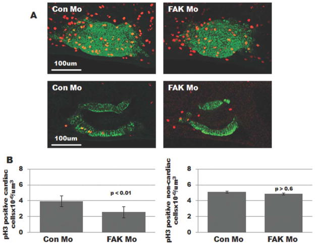
FIG. 5.
Myocyte mitosis is attenuated in FAK morphant heart tubes. A: Whole mount immunohistochemical staining for tropomyosin (green) and phospho-Histone H3 (red) was performed on prelooped (Stage 32) embryos that were injected with either Con Mo (left) or FAK Mo (right) at the one-cell stage. Images represent 3D reconstructions of confocal z-stack sections (top panels) or a single optical section (bottom panels). B: Total number of pH3 positive myocytes and noncardiac cells were counted in each optical section of Con Mo- and FAK Mo-injected embryos (19 embryos were analyzed per condition, collected from at least three separate experiments).

