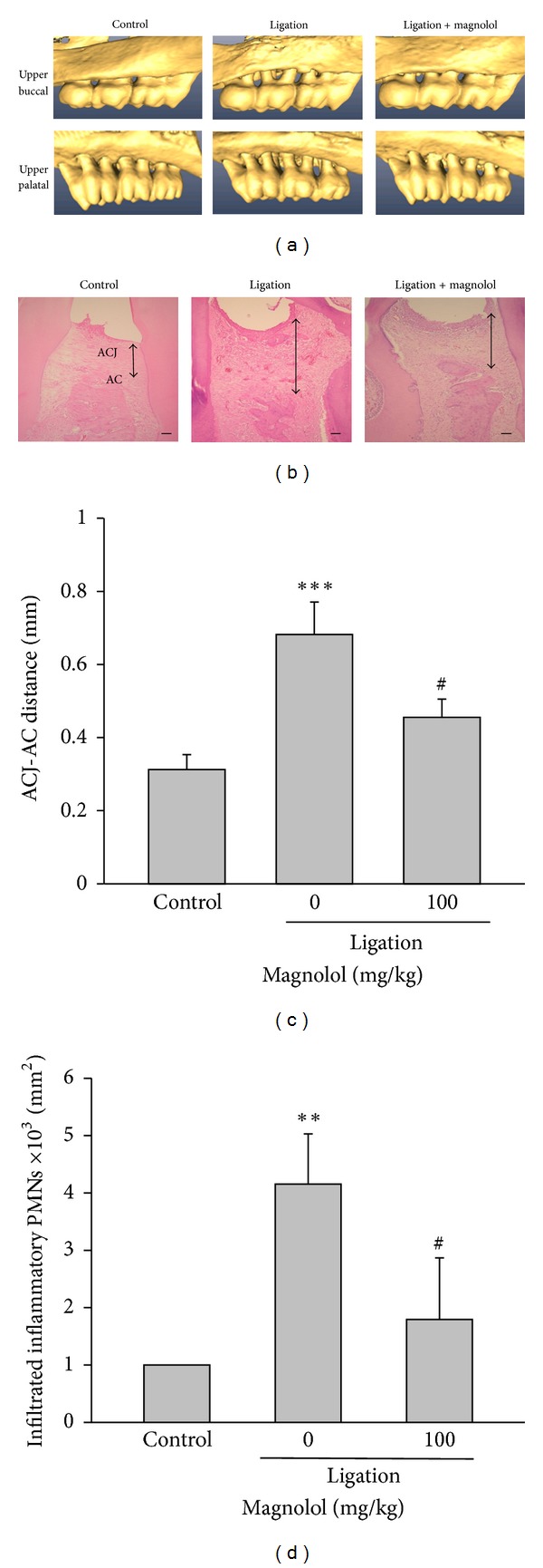
Figure 1.

Effect of magnolol on alveolar bone loss and tissue damage. (a) Reconstructed three-dimensional micro-CT images (buccal and palatal view) of the maxilla second molars buccal (upper panel) and palatal (lower panel) alveolar bone level in nonligation (control), ligation, and ligation + magnolol (100 mg/kg) groups at the experimental day 8. (b) Histological observation (H&E stain) of maxillary intermolar tissue in various groups (scale bar = 100 μm). (c) The degree of alveolar bone loss was evaluated by measuring the ACJ-AC distance at the buccal furcation site of the maxilla second molar from the reconstructed micro-CT images. (d) Quantitative analysis of the total number of inflammatory infiltrated cells in maxillary intermolar gingivomucosal tissue was performed by accounting the number of polymorphonuclear cells (PMNs). Data are expressed as the mean ± SD (n = 5). ***P < 0.001 versus control; # P < 0.05, ## P < 0.01 versus ligation group.
