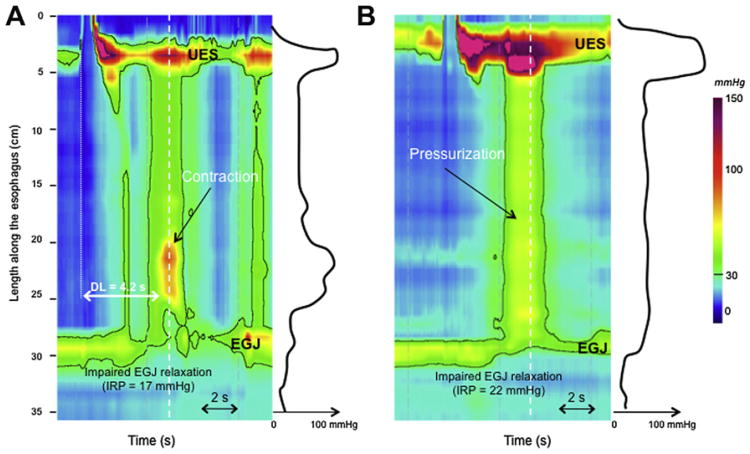
Fig. 2.
Spastic (type III) achalasia is characterized as impaired EGJ relaxation associated with at least 20% of premature contractions (A). The premature contraction exhibits a reduced DL (<4.5 s). EGJ relaxation is assesses using the IRP. Simultaneous (premature) contractions might be differentiated from pressurization (B) using the spatial pressure variation plot represented on the right of each EPT plots. Each spatial pressure variation pressure plot was obtained at the time, identified by the white dashed line. In the instance of an esophageal contraction (A), pressure variations are obvious along the esophageal body. In instances of pressurization (B), intraesophageal pressure did not vary between UES and the EGJ. (B) This corresponds to type II achalasia (achalasia with compression).

