
Figure 7.
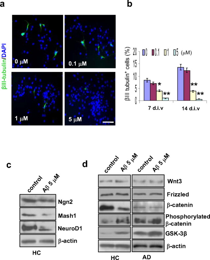
Effect of Aβ treatment on neurogenesis and β-catenin signaling. HC GPC neurospheres were treated with different concentrations of Aβ1–42 peptide for 2 d. Then, the Aβ was removed, and the cultures were tested after either 7 or 14 d in differentiation medium. a, After 7 d of differentiation, immunostaining revealed a dose-related reduction in the percentage of neurons (βIII tubulin+) among the Aβ-treated HC GPC progeny (labeled with DAPI). Scale bar, 50 μm. b, Quantification of immunoreactive structures after 7 and 14 d of differentiation showed that the percentage of new neurons among HC GPC progeny decreased significantly with ≥1.0 μm Aβ1–42 exposure compared with controls (0 μm Aβ1–42). c, A representative Western blot of HC GPC progeny treated with 5 μm Aβ1–42 revealed reduced levels of proneural gene products Ngn2, Mash1, and NeuroD1 compared with control HC progeny. d, HC and AD GPCs were treated with 5 μm Aβ1–42 for 2 d, and the culture was extended to 7 d in Aβ-free differentiation medium. Western blot showed a reduction in nonphosphorylated β-catenin and increases in both GSK-3β and phosphorylated β-catenin in Aβ-treated HC GPC progeny compared with control progeny. In AD GPC progeny, the blot did not show any significant change in the levels of nonphosphorylated or phosphorylated β-catenin between the Aβ-treated and control progeny; however, the Aβ-treated AD GPC progeny did exhibit a significant increase in GSK-3β expression compared with control AD progeny. No significant differences in either Wnt3 or frizzled levels were observed between Aβ-treated and control progeny from HC or AD GPCs (n = 3 per group).