Fig. 4.
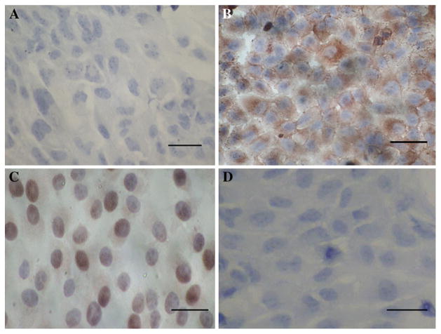
The immunocytochemistry results of C-MYC in HFE-PC and HFE-Myc cells (×400). a There was no positive signal in HFE -PC cell. Bar 20 μm. b The brown positive signals were mainly distributed in cytoplasm of HFE-Myc cells. The results showed that there was expression of C-MYC gene in HFE-Myc cell line. Bar 20 μm. c The brown positive signals were mainly distributed in nucleus and cytoplasm of positive control cells. Bar 20 μm. d There was no positive signal in HFE-Myc cells which were immunostained using PBS instead of C-MYC antibody as negative control. Bar 20 μm. (Color figure online)

