
Figure 6. Recirculating Kaede-red CD3+ CD4+ CD44hi T cells up-regulate CD40L and secrete IL-2.
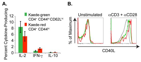
Shaved abdominal skin of Kaede transgenic mice was exposed to 420 nm light for 5 minutes. After 24 h, draining LNs were isolated and (a) recovered cells were stimulated for 4 h with plate-bound anti-CD3 and soluble anti-CD28. Cytokine secretion by Kaede-green CD3+ CD4+ CD44hi CD62Lhi memory T cells and Kaede-red CD3+ CD4+ CD44hi memory T cells was analyzed by flow cytometry. Data are pooled from 3 experiments with 9 mice total for each cytokine. (b) Or, CD4+ T cells were purified from draining LNs and stimulated for 2 h with PMA and ionomycin, or left untreated. Then, surface CD40L expression was analyzed on Kaede-green CD44lo naïve T cells (gray lines), Kaede-green CD44hi memory T cells (green lines) or Kaede-red CD44hi memory T cells (red lines). Data are representative of 2 experiments with 6 mice total.