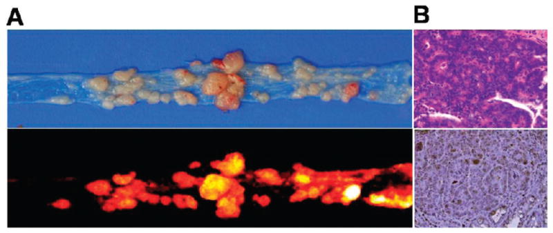
Figure 4.

(A) Upper: photo image of colon tumors from an A/J mouse treated with AOM. Lower: NIR fluorescence image of colon tumors after intravenous injection of the NS. (B) Immunohistology analysis of colon tumors. Upper: H&E stain; lower: IHC of MMP-9 protein expression in a colon tumor section counterstained with primary antibody for MMP-9.
