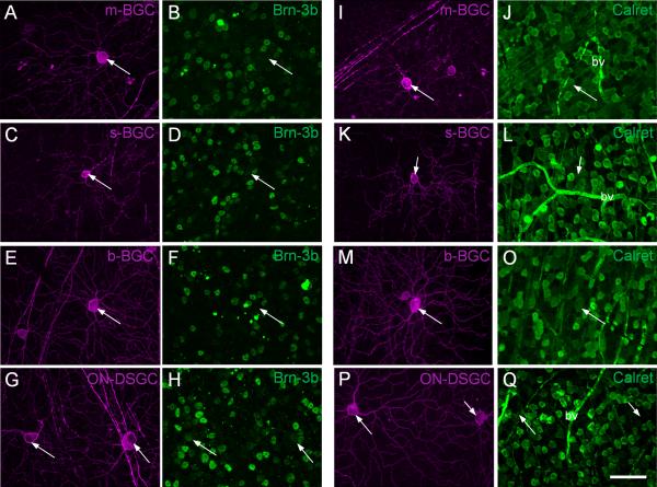
Fig. 7.
Immunostaining of PCP2-BGCs for Brn-3b and calretinin. A-B: The nucleus of m-BGC (A, arrow) was negative for Brn-3b (B, arrow). C-D: The nucleus of the s-BGC (C, arrow) was sometimes weakly stained with the antibody against Brn-3b (D, arrow). E-F: b-BGCs were negative for Brn-3b (arrow). G-H: On-DSGCs were positive for Brn-3b (arrows). I-O: The somas of m-BGC (I-J, arrow), s-BGC (K-L, arrow), and b-BGC (M-O, arrow) were positive for calretinin. P-Q: The somas of the two putative On-DSGCs (arrows) were negative for calretinin. bv - blood vessel. Scale bar = 50 μm in Q (applies to all panels).

