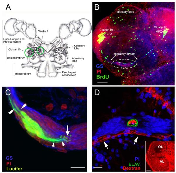
Figure 1.
(A) Diagram of the eureptantian (crayfish, lobster) brain including the optic ganglia, and showing the locations of the proto-, trito- and deutocerebral neuropils. The soma clusters 9 and 10 (circles), locations of neurogenesis in the adult brain, flank two prominent neuropil regions of the deutocerebrum, the olfactory and accessory lobes. (Names of brain areas are according to Sandeman et al., 1992.) (B) Left side of the brain of Procambarus clarkii labeled immunocytochemically for the S-phase marker BrdU (green). Labeled cells are found in the lateral proliferation zone (LPZ) contiguous with Cluster 10 and in the medial proliferation zone (MPZ) near Cluster 9. The two zones are linked by a chain of cells in the migratory stream, labeled immunocytochemically for glutamine synthetase (GS; blue). These streams originate in the oval region ‘niche’ (dotted circle) containing cells labeled with the nuclear marker propidium iodide (PI, red). (C) Several niche cells are labeled by intracellular injection of Lucifer yellow. Each of these has a short process (arrowheads) projecting to the vascular cavity (arrow) and longer fibers (double arrowheads) that fasciculate to form the tracts projecting to the LPZ and MPZ, along which the daughters of the niche cells (2nd-generation neuronal precursors) migrate (the ‘streams’). Blue, GS; red, propidium iodide (PI). (D) The vascular connection of the cavity in the center of the glial soma cluster was demonstrated by injecting a dextran dye into the dorsal artery. The cavity, outlined in green by its reactivity to an antibody to ELAV, contains the dextran dye (red), which is also contained within a larger blood vessel that runs along beneath the niche. PI (blue) labeling of nuclei in the niche cells is also shown. Inset: dextran-filled vasculature in the olfactory (OL) and accessory (AL) lobes on the left side of the brain. Scale bars: B, 100 μm; C and D, 20 μm; inset in D, 100 μm. [C and D from Sullivan et al., 2007a].

