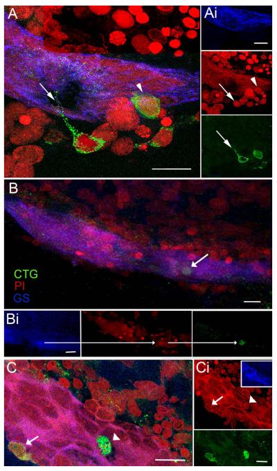
Figure 6.
Cells circulating in the hemolymph are attracted to the niche in vitro. CellTracker™ Green (CTG)-labeled hemocytes are found in the vascular cavity and in, and on, the neurogenic niche. (A) Niche on a desheathed brain co-cultured with CTG-labeled cells extracted from the hemolymph: merged confocal fluorescent channels of stacked images. Several CTG-labeled cells reside just outside the niche, one with a long process extending into the niche (arrow), and another that is inserting on the outer margin of the niche (arrowhead). (Ai) Separate channels: GS outlining the niche (top); PI revealing cell nuclei, arrow/arrowhead pointing to the respective CTG-labeled cells in A (middle); and CTG-labeled cells with arrow pointing to the fine process from the CTG-labeled cell (bottom). (B) Projection of stacked images from a more dorsal region of the neurogenic niche than in (A) reveals a CTG-labeled cell just below the surface of the niche. (Bi) Separate confocal channels of the region in (B) with arrows pointing to the same CTG-filled cell also labeled for GS (left); PI, revealing cell nucleus (middle); and CTG-labeling (right). (C) In another example, a CTG-labeled cell (arrowhead) resides in the cavity and a second CTG-labeled cell (arrow) is embedded in the outer edge of the niche. (Ci) PI labeling of cell nuclei with arrowhead and arrow pointing to the corresponding nuclei with CTG-labeling in C. Insert, GS labeling of the niche. Bottom, separate channel, CTG-labeled cells. Scale bars: A, 20 μm; Ai, 10 μm; B, Bi, C and Ci, 20 μm.

