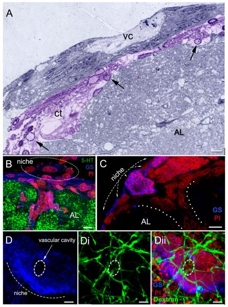
Figure 8.
The relationship between the neurogenic niche and vascular tissues. (A) Semi-thin section stained with toluidine blue, showing the niche and central vascular cavity (vc). The connective tissue (ct) below the niche (colorized purple) has many cells (arrows) with features resembling hemocytes. (B, C) Sagittal sections through the brain. These images show the niche lying on the ventral surface of the accessory lobe (AL), which is labeled immunocytochemically for serotonin (5-HT; green) in B. A blood vessel (dotted lines in C) emerges from the accessory lobe, Some cells within the blood vessel and others forming a layer between the niche and accessory lobe (B) are immunoreactive for glutamine synthetase, as are the niche cells. (D) The dorsal sinus in adult crayfish (15-20 carapace length) was injected with fluorescently-labeled dextran, which rapidly filled the brain vasculature. Fine blood vessels (dextran, green) associated with the niche are revealed on the ventral surface of the niche (Di, Dii), with some of these infiltrating the edge of the vascular cavity (broken line circle). Scale bars: A and C,10 μm; B, 6 μm; D, 20 μm. [A from Chaves da Silva et al., 2012]

