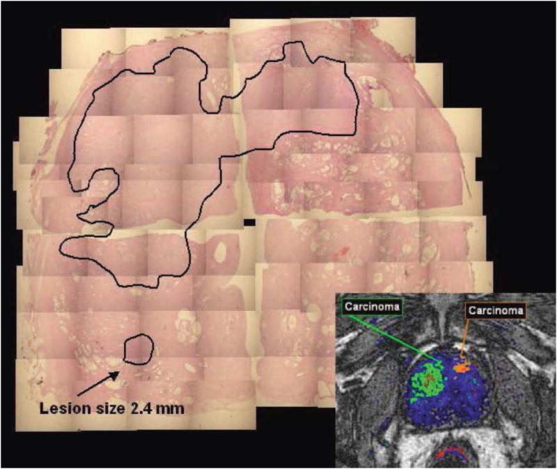
Figure 3.
Two large lesions were detected by DCE-MRI. The lesion in the dorsal part of the right lobe of the prostate being smaller than 3 mm and containing less than 30% of cancer cells was not detected by DCE-MRI. (Figure 6 in SCHMUECKING et al, Int. J. Radiat. Biol., Vol. 85, No. 9, September 2009, pp. 814–824, pending on copy right permission from the journal)

