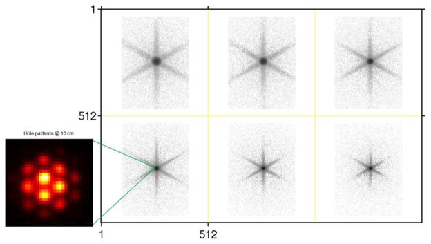
Fig. 1.

Measured I-131 point source images at six different distances between the front surface of the collimator and the object plane. Top row: 25, 20, 15 cm, bottom row: 10, 5, 2 cm. Zoomed point source image at 10 cm shows hole pattern due to high-energy collimator. The image intensity is presented on a logarithmic grey scale except for the zoomed image.
