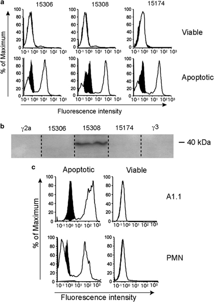
Figure 1.
Anti-LPS antibodies crossreact with multiple epitopes of apoptotic cells. (a) Flow cytometric profiles of MUTU I BL cells triggered into apoptosis by ionomycin and stained using the indicated anti-LPS mAbs (white histograms), which were detected by fluorescein isothiocyanate (FITC)-conjugated goat anti-mouse immunoglobulin G (IgG); isotype control binding (γ2a for mAbs 15174 and 15306, γ3 for mAb 15308) is indicated by the black histograms. (b) Immunoblotted SDS-PAGE of MUTU I lysates probed with the indicated anti-LPS mAbs or isotype controls demonstrating 40 kDa species specified by mAb 15308 but not 15174 or 15306. (c) Flow cytometric profiles of A1.1 hybridoma cells (upper panels) and human neutrophils (lower panels) after induction of apoptosis by ionomycin or serum deprivation, respectively. Cells were stained with mAb 15308 detected as above; isotype control binding shown in black. Apoptotic and viable cells were discriminated by light scatter as described6

