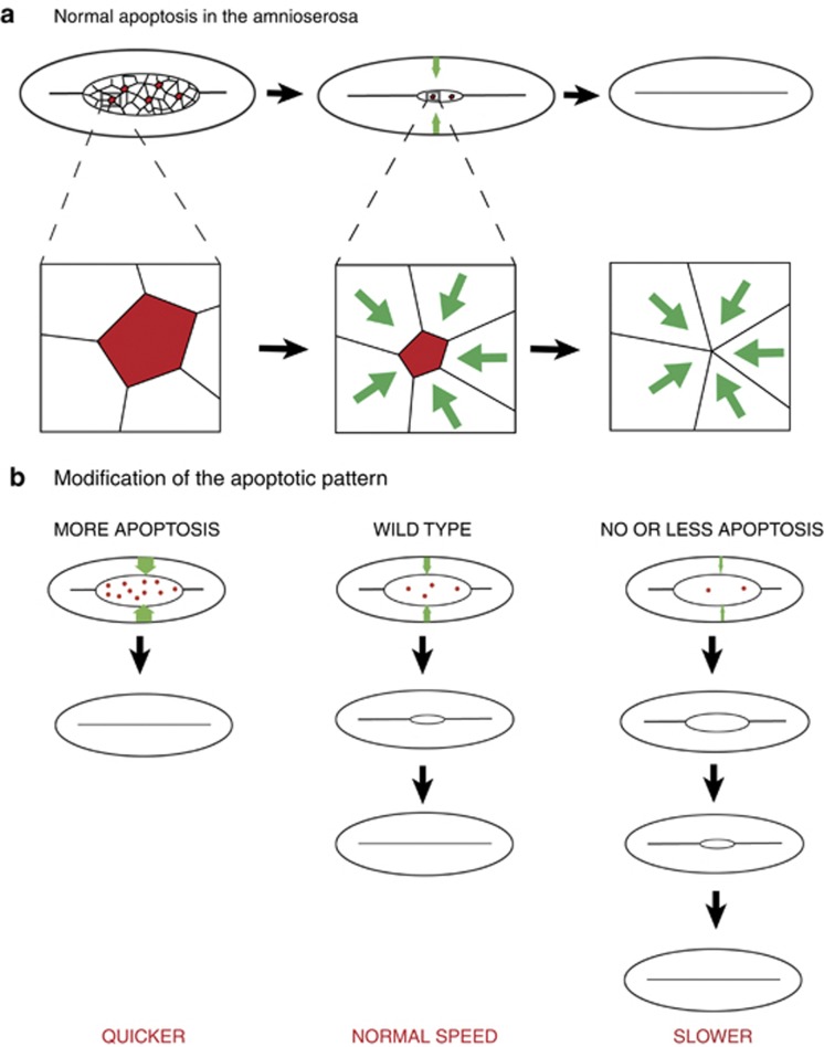
Figure 2.
Apoptosis acting as a pulling force. (a; up) Schematic representation of a dorsal view of a Drosophila embryo during dorsal closure. Apoptotic cells are indicated in red (in the amnioserosa), the force generated by apoptosis in the lateral epidermis is represented by the green arrows. Successive stages of dorsal closure are represented. (a; down) Representation of an apoptotic cells and its closest neighbors at higher magnification. The progressive stretching of the neighboring cells is proposed to be the origin of the pulling force generated by apoptosis on the surrounding tissue.(b) Dorsal closure defects when apoptosis pattern is modified. Dorsal closure can be either accelerated when apoptosis is promoted (left), or slowed down when apoptosis is inhibited (right)

