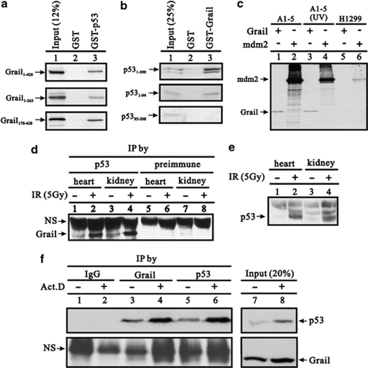
Figure 1.
Interaction between Grail and p53 in vitro and in vivo. (a and b) Full-length Grail or Grail fragments (a) were translated in vitro and incubated with bead-bound GST or GST-p53. (b) Full-length p53 or p53 fragments were translated in vitro and incubated with bead-bound GST or GST–Grail. Bound proteins were eluted, separated by SDS-PAGE, and subjected to autoradiography. (c) A1–5 and H1299 cell lysates incubated at 37 °C or after treatment with 10 J/m2 UV were subjected to IP with pAb421 and Protein A Sepharose beads. The pulled-down immunocomplexes were incubated with 35S-labeled Grail or Mdm2 as indicated, resolved by SDS-PAGE, and visualized by autoradiography. (d–e) Mouse tissue extracts were prepared from mice treated either with or without IR, followed by IP with a p53 antibody and western blot analysis with a Grail antibody or pre-immune serum. (e) Input of tissues served as the IP control. (f) A549 cell extracts were prepared from cells treated either with or without Act D followed by IP with p53, Grail, or IgG antibody and western blot analysis with Grail or p53 antibody. (NS, non-specific band)

