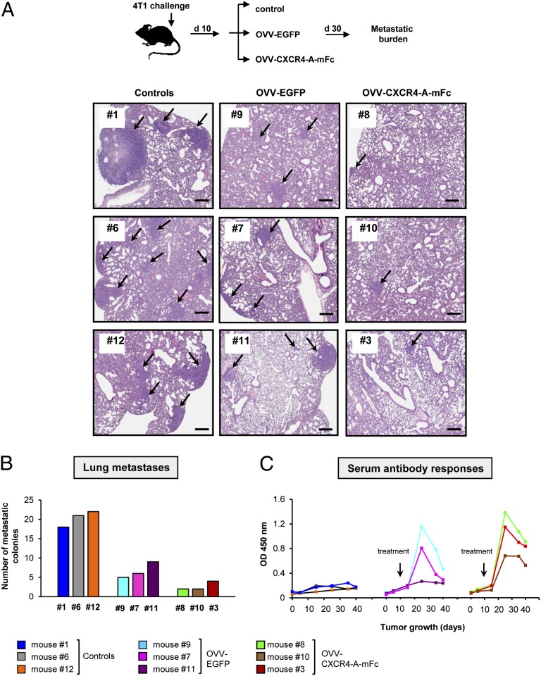
Fig. 4.
Evaluation of lung metastases and antibody responses in mice after onvolytic virotherapy. (A) The oncolytic virotherapy was initiated once the primary tumor reached ∼150 mm3 (n = 3 mice per group). Metastatic growth in control and treatment groups were monitored by bioluminescence for 30 d until killing was performed at the time of excessive tumor burden in control mice (Upper), after which lung metastases were assessed by histology on formalin-fixed and H&E-stained sections (Lower). (Scale bars, 300 μm.) (B) Numbers of metastatic colonies per section were presented in individual mice. (C) Sera collected before tumor challenge, at the time of orthotopic 4T1 challenge, at the time of treatment, and every 10 d until killing were analyzed for the presence of antitumor antibody responses by ELISA using wells coated with 47-LDA mimotope of ALCAM/CD166. All samples were analyzed in triplicates with serum dilution of 1:100.

