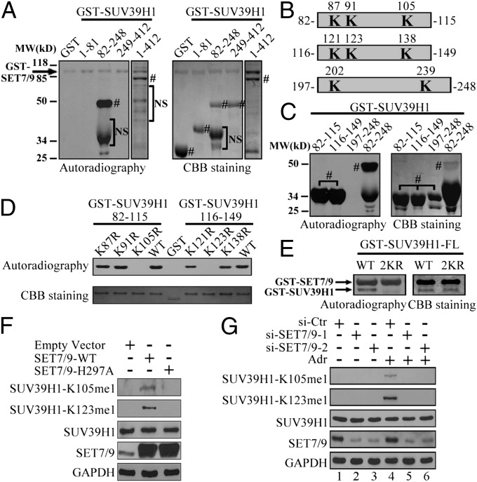
Fig. 2.
SET7/9 methylates SUV39H1 in vitro and in vivo. (A) GST–SUV39H1 FL or fragments were incubated with SET7/9 and 3H–SAM for 1 h at 30 °C. The samples were subsequently separated by SDS/PAGE, stained by Coomassie brilliant blue (CBB), or exposed by autoradiography. The arrow indicates automethylation of GST-SET7/9. # represents specific protein bands; NS refers to nonspecific bands. (B) A schematic diagram of the subfragments of SUV39H1. The localization of lysines is in bold. (C) GST–SUV39H1 (82–115 aa) or three subfragments mentioned in B were catalyzed by SET7/9, and autoradiography or CBB staining was performed as indicated in A. # represents specific protein bands. (D) Indicated GST–SUV39H1 fragments with or without individual lysine mutations were catalyzed by SET7/9 and analyzed by autoradiography or CBB staining. (E) FL GST–SUV39H1–WT or –2KR was catalyzed by SET7/9 and analyzed as described in A. (F) Empty vector, flag–SET7/9–WT, or –H297A was transfected into H1299 cells for 48 h. Western blotting was then performed with indicated antibodies. (G) siRNA control (si-Ctr) or two SET7/9 siRNA fragments were transfected into H1299 cells for 48 h with or without 1 μM Adr for the last 24 h. Western blotting was subsequently performed by using the indicated antibodies.

