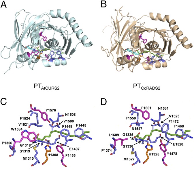
Fig. 4.
Homology models of PTCcRADS2 and PTAtCURS2. Cartoon views of the homology models and stick representation of the amino acids lining the cyclization chambers of (A and C) PTAtCURS2 and (B and D) PTCcRADS2. The side chains of residues discussed in the text are shown as golden (catalytic histidine), magenta (residues implicated in differentiating substrate orientation), and blue (other significant residues) sticks. Green sticks show the substrate analog palmitic acid resident in the PTNSAS structure 3HRQ (10).

