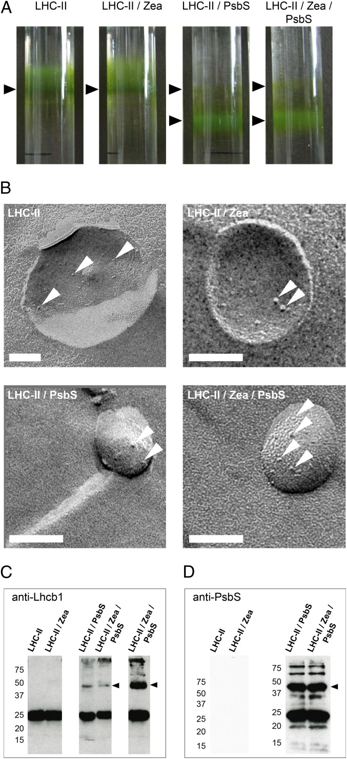
Fig. 2.
Proteoliposomes for in vitro investigation of qE constituents. (A) Proteoliposome bands on sucrose density gradients (arrowheads). The band above the opaque LHC-II/PsbS proteoliposome fraction contained very small liposomes with little incorporated protein and was not used further. (B) Freeze-fracture electron micrographs of proteoliposomes (protein complexes are indicated by arrowheads). (Scale bars: 100 nm.) Western blots of proteoliposomes with Lhcb1 (C) or PsbS (D) antibodies. Lanes labeled LHC-II/Zea/PsbS in C are from the same sample loaded at different concentrations. Arrowheads indicate a heterodimer consisting of LHC-II and PsbS (Left, molecular masses in kilodaltons).

