Figure 4.

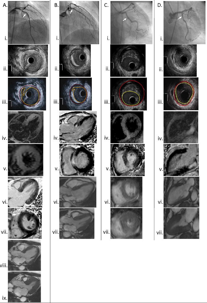
Co-localization of angiographic, IVUS and CMR images in patients with different constellations of findings. The site of IVUS imaging is marked on each angiogram.
A: Plaque rupture with increased T2 signal and absent LGE. Left coronary angiogram (i) with corresponding IVUS showing plaque rupture in the mid LAD (ii-iii). T2 signal hyperintensity is noted to involve the mid anterior and mid and apical anterior septal walls (iv-v). No LGE is noted within the myocardium in 2 planes (vi-vii). Normal LV function (viii-ix).
B: Plaque rupture with LGE. Angiogram of the left coronary artery (i) with corresponding IVUS showing plaque rupture in the proximal LAD (ii-iii). The site of the IVUS images is marked with an arrow. Subendocardial to transmural LGE involving the basal anteroseptal wall in shown in 3-chamber and short axis views (iv-v). T2-weighted imaging was not performed in this patient. Normal LV function (vi-vii).
C: Ischemic-type LGE without plaque rupture on IVUS. Left coronary angiogram (i). IVUS of the LCX showing mixed-fibrofatty plaque without rupture (ii-iii). The LAD and LCX were imaged with IVUS in this case. T2 signal hyperintensity of the mid inferior, mid inferoseptal and adjacent right ventricular walls near the right ventricular insertion (iv). Transmural LGE involving subsegments of the same region (v). Normal LV function (vi-vii)
D: Non-ischemic type LGE with minimal atherosclerosis on IVUS. Left coronary angiogram (i). Normal IVUS of the LCX (ii-iii). T2 signal hyperintensity of the basal inferior/inferolateral wall (iv). Focal area of mid-myocardial LGE involving the same region. (v) Normal LV function (vi-vii).
