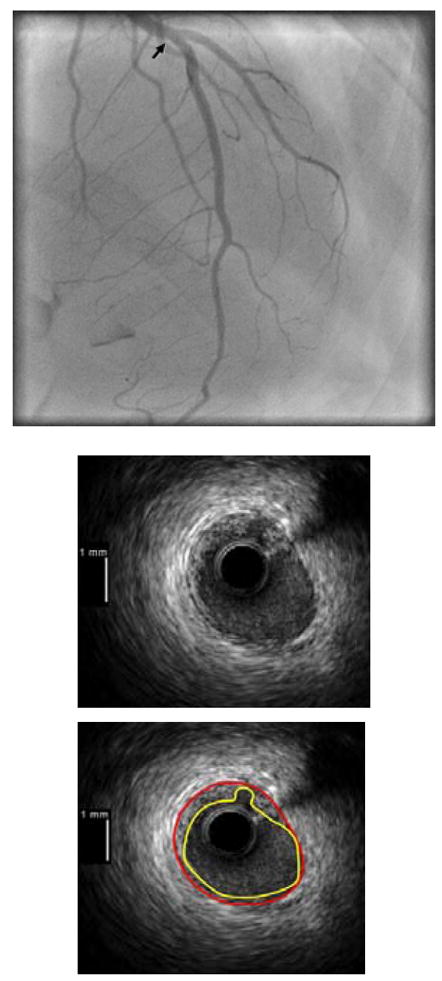
Figure 5.

Angiographic and IVUS images from a patient with tako-tsubo cardiomyopathy and plaque ulceration. Angiogram shown in the top panel with arrow marking the site of the IVUS image shown in the lower panels, with and without contours outlining the intimal border (yellow) and external elastic lamina (red). Note that the LAD is a large vessel which wraps around the LV apex; this was also true of the other case of tako-tsubo cardiomyopathy with plaque ulceration in the left main coronary artery (not shown).
