Fig. 3.
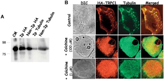
Immunoprecipitation and co-localization of TRPC1 with tubulin. A: Co-immunoprecipitation of TRPC1 with the cytoskeletal protein tubulin. Crude membranes prepared from ARPE cells transiently expressing HA-TRPC1 were solubilized and immunoprecipitation was performed as described in experimental procedures. Proteins in the IP were detected using SDS PAGE and immunoblotting. Antibodies used for IP and IB are indicated in the figure. Control IPs is shown by crude membranes of HA-TRPC1 expressing ARPE cells. B: Co-localization of HA-TRPC1 with endogenous tubulin proteins in ARPE cells. Anti-HA antibody, anti tubulin antibody, and FITC or rhodamine-conjugated secondary antibodies were used to detect transiently expressed HA-TRPC1 and endogenous tubulin in ARPE cells. Overlay of the images is shown in the right panel (yellow signal). Confocal pictures of the transiently expressing HA-TRPC1 and tubulin proteins in ARPE cells treated with colchicine (lower panels). Arrows in the images show the protein localization.

