Fig. 4.
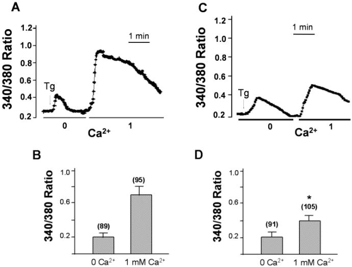
Colchicine treatment decrease thapsigargin stimulated TRPC1 activity. Ca2+ influx were measured in Tg-stimulated cells in a Ca2+-free buffer, followed by addition of 1 mM Ca2+ to the medium. A, C: Fluorescence traces in either control ARPE cells (A) or colchine-treated ARPE cells (B) stimulated with Tg. B, D: Bar graphs showing the relative Ca2+ influx in the absence and in the presence of extracellular Ca2+. “*” indicate values that are significantly different from that of the respective control condition (P < 0.02, number of cells is indicated in each case). Internal Ca2+ release and basal Ca2+ influx were not altered by colchine treatment.

