
Fig. 6. Overexpression of TRPC1 decreases proteins required for apoptotic pathway.
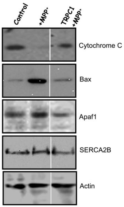
Shown is a Western blot performed on a mitochondrial membrane fraction. 25 μg of the membrane fraction was resolved on 4–20% SDS gel and probed with different antibodies. Bound cytochrome c was assayed using anti-cytochrome c antibody (top panel). Bax and SERCA2B proteins were identified using anti-Bax or anti-SERCA2B antibodies. Similarly, Apaf-1 and actin antibodies were used to detect proteins from the total cell lysates, respectively. Details about the antibodies and methods are described under “Experimental Procedures.”