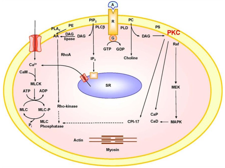
Figure 1.
Mechanisms of VSM contraction. The interaction of an agonist (A) such as phenylephrine with its specific α-adrenergic receptor (R) activates phospholipase C (PLCβ) and stimulates the hydrolysis of phosphatidylinositol 4,5-bisphosphate (PIP2) into inositol-1,4,5-trisphosphate (IP3) and diacylglycerol (DAG). IP3 stimulates Ca2+ release from the sarcoplasmic reticulum (SR). Agonists also stimulate Ca2+ influx through Ca2+ channels. Ca2+ binds calmodulin (CAM), activates MLC kinase (MLCK), causes MLC phosphorylation, and initiates VSM contraction. DAG activates PKC. PKC-induced phosphorylation of CPI-17, inhibits MLC phosphatase and increases MLC phosphorylation and VSM contraction. PKC-induced phosphorylation of the actin-binding protein calponin (CaP) allows more actin to bind myosin and enhances contraction. PKC may also activate a protein kinase cascade involving Raf, MAPK kinase (MEK) and MAPK, leading to phosphorylation of the actin-binding protein caldesmon (CaD) and enhanced contraction. Activation of RhoA/Rho-kinase inhibits MLC phosphatase and further enhances the Ca2+ sensitivity of contractile proteins. AA, arachidonic acid; G, heterotrimeric GTP-binding protein; PC, phosphatidylcholine; PE, phosphatidylethanolamine; PLD, phopholipase D; PS, phosphatidylserine. Dashed line indicates inhibition.

