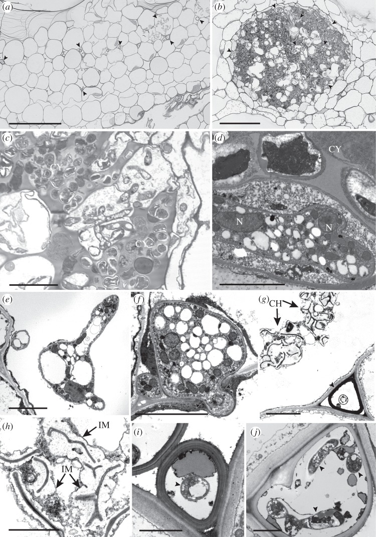
Figure 1.
(a–c) Light and (d–j) transmission electron micrographs of fungal endophytes in (a,g,h) Folioceros (MA33) and in (b–f) Anthoceros (MA29). (a–c) Fungal hyphae occur either (a) scattered in the central region of the thallus (arrowed) or (b) in close association with cyanobacterial colonies (CY; arrowed, enlarged in c). (d) Multi-nucleate hypha (N, nucleus) in cell adjacent to a cyanobacterial colony (CY). (e,f) Intracellular hyphae; (e) branched hypha and (f) hypha bridging the walls (arrowed) of two adjacent host cells. (g) Thick-walled fungal structure in mucilage-filled intercellular space (arrowed) adjacent to intracellular collapsed hyphae (CH). (h) Detail of collapsed intracellular hyphae, note the extensive interfacial matrix (IM). (g,j) Intercellular thick-walled fungal structures with internal thin-walled hyphae (arrowed). Scale bars: (a,b) 100 µm, (c) 20 µm, (d–g) 5 µm, (h–j) 2 µm.

