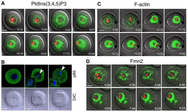
Fig. 1.

Constitutive synthesis of PtdIns(3,4,5)P3 during meiotic maturation of mouse oocytes. GV oocytes were injected with mRNAs encoding PH-Akt–GFP, EGFP–Lifeact and EGFP–Fmn2 and imaged by time-lapse confocal microscopy during meiotic maturation to document the localization of (A) PtdIns(3,4,5)P3, (C) F-actin and (D) Fmn2. (B) Fixed GV oocytes were stained with an antibody against p85 to document the presence of PI3K in the vicinity of chromosomes (arrowheads). DNA (red or blue) was labeled with Hoechst 33258. Images in A, C and D were taken from individual sections of confocal images at the indicated time points (hr:min). Scale bars: 20 µm.
