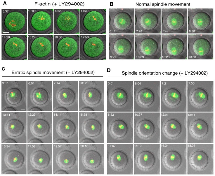
Fig. 2.
Blockage of PtdIns(3,4,5)P3 synthesis disrupted dynamic F-actin assembly and spindle translocation to the cortex. GV oocytes were matured in vitro and imaged by time-lapse confocal microscopy as described in Fig. 1. After treatment with LY294002 (20 µM) F-actin (A) diffused throughout the cytoplasm and the normal translocation of the spindle from the center to cortex (B) became erratic (C) or occurred with frequent changes in spindle orientation without net displacement (D). F-actin in normal controls was as in Fig. 1C. Scale bars: 20 µm.

