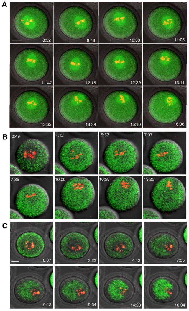
Fig. 4.

Absence of MATER protein impaired PtdIns(3,4,5)P3 synthesis and spindle translocation. GV oocytes from Mater−/− females and the wild-type littermates were matured in the same droplet of medium. The zona pellucida was removed from mutant or normal oocytes. (A,B) Time-lapse confocal microscopy was performed as described in Fig. 1 to document that meiotic spindles move erratically in mutant oocytes (A) compared with normal oocytes (supplementary material Fig. S4) , and that (B) PtdIns(3,4,5)P3 was diffusely present in oocytes lacking MATER but not in normal control oocytes (supplementary material Fig. S5). (C) Symmetric F-actin assembly was lost in Mater−/− oocytes but not in normal control oocytes (supplementary material Fig. S8). However, the asymmetric F-actin cloud was visible when chromosomes approached to the cortex. Scale bars: 20 µm.
