Fig. 3.
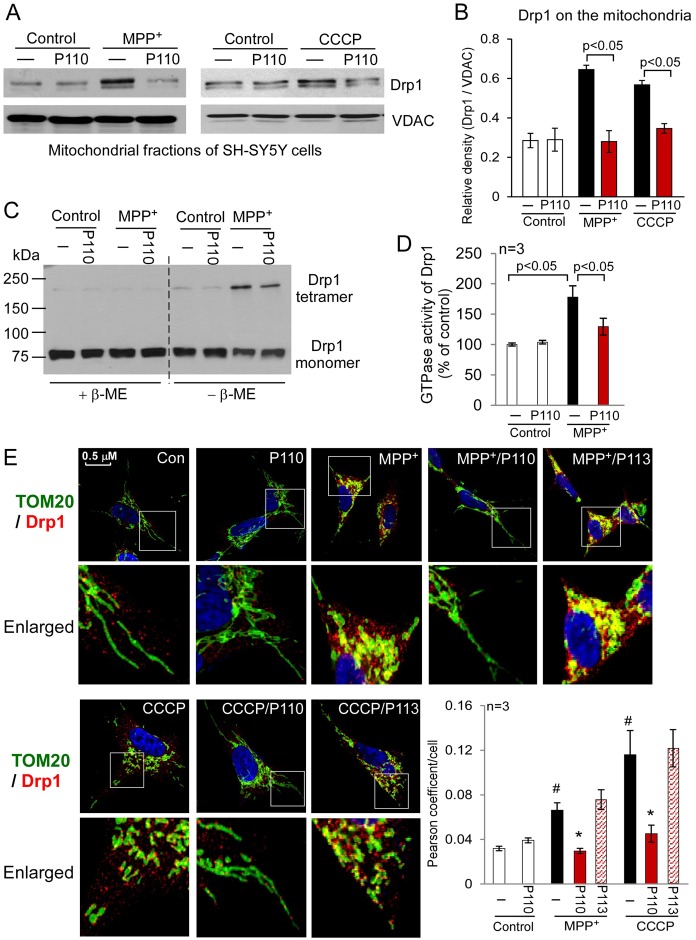
Peptide P110 specifically inhibited Drp1 interaction with the mitochondria in cultured SH-SY5Y neuronal cells exposed to mitochondrial stressors. Cultured human SH-SY5Y neuronal cells were treated with peptide P110 (1 µM) for 30 minutes prior to a 1 hour incubation in the absence or presence of MPP+ (2 mM) or CCCP (10 µM). (A) Western blot analysis of mitochondrial fractions was determined by the indicated antibodies. VDAC was used as a loading control. (B) Quantification of the levels of Drp1 is provided in a histogram. Data are expressed as means ± s.e. of three independent experiments. (C) Total lysates of SH-SY5Y cells from the indicated groups were subjected to western blot analysis using reducing or non-reducing gels, and monomeric (∼75 kDa) and tetrameric (∼200 kDa) Drp1 levels were determined. (D) The GTPase activity of immunoprecipitated Drp1 from cultured cells was expressed as the mean ± s.e. of three independent experiments. (E) Confocal microscopy of stained SH-SY5Y cells with anti-Drp1 (1:500 dilution) and anti-Tom20 (a marker of mitochondria, 1:500 dilution) antibodies following incubation with either MPP+ (2 mM for 1 hour) or CCCP (5 µM for 30 min). Lower panels show enlarged areas of the white boxes in the above panels. Scale bar: 0.5 µm. Pearson's coefficient/cell (Drp1/Tom20 co-localization) was determined using confocal microscopy (Fluoview FV100, Olympus) and is provided as a histogram, as mean ± s.e. of three independent experiments. (#P<0.05 versus control cells; *P<0.05 versus cells treated with MPP+ or CCCP.)

