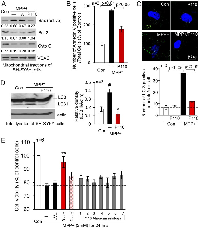
Fig. 6.
Treatment with P110 reduced cell death induced by mitochondrial stressors. (A) SH-SY5Y neuronal cells were treated with P110 followed by exposure to MPP+ (2 mM for 1 hour). The active form of Bax (NT-Bax), cytochrome c and Bcl-2 on the mitochondria were determined by western blot analysis with the indicated antibodies. Shown are representative data of three independent experiments. (B) Apoptosis was determined using annexin V staining 8 hours after MPP+ exposure. The number of apoptotic cells was expressed as mean ± s.e. of percentage relative to total number of cells from three independent experiments. (C) Cultured SH-SY5Y cells were stained with anti-LC3 antibodies (green); punctate LC3 staining is an indicator of autophagy. Nuclei were stained with Hoechst dye. Upper panel: representative images. Lower panel: histogram depicting the number of LC3-positive puncta/cell in the indicated groups. The data are presented as the means ± s.e. of three independent experiments, scored by an observer blinded to the experimental conditions. (D) Western blot analysis of LC3 I versus LC3 II was determined with anti-LC3 antibodies. Histogram: the data are expressed as means ± s.e. of three independent experiments. (E) Cell viability was measured by MTT assay. SH-SY5Y cells were treated with MPP+ (2 mM for 24 hours) following treatment with TAT, P110, P113 or P110 Ala-scan analogs (1–7, 1 µM each). #P<0.05 versus control group; *P<0.05, **P<0.01 versus MPP+-treated cells. Con, control.

