Figure 3.
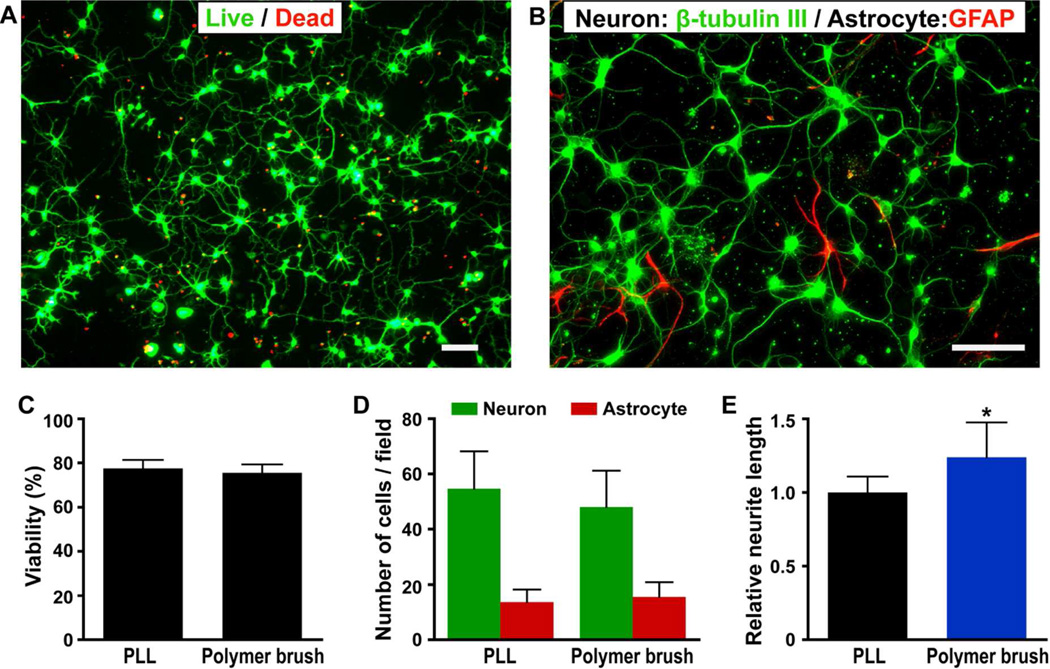
Photographs of mouse hippocampal neuronal cells cultured on polymer brush modified silicon surfaces and data comparison with those on poly-L-lysine coated glass slides. A) LIVE/DEAD viability/cytotoxicity assay of hippocampal cells on polymer brushes (scale bar is 50 µm). Cells were cultured for three days and observed with fluorescent micrographs of live (calcein AM, green) and dead (ethidium homodimer-1, red). B) Immunocytochemistry demonstrates that neurons (β-III-tubulin-positive cells, green) outnumber astrocytes (GFAP-positive cells, red) on the surface. (Scale bar is 50 µm). C) Viability of cells on polymer brush is comparable to that of PLL coated glass slides. D) Similar number of neurons and astrocytes were observed on polymer brushes and the PLL control surfaces. E) Neurons on polymer brush surface exhibit longer neurites as compared to those on the PLL coated glass slides.

