
Figure 1.
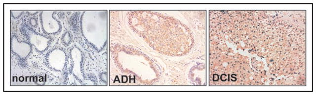
Analyses of VEGFR-3 expression in human breast cancer specimens. Human breast samples staining with VEGFR-3 9D9F9 antibody. Left panel normal tissue, center hyperplasia ADH measured as 2+, right—DCIS staining measured as 2+. Diaminobenzidine (DAB) was used as the chromogen, and the slides were counter-stained with hematoxylin.