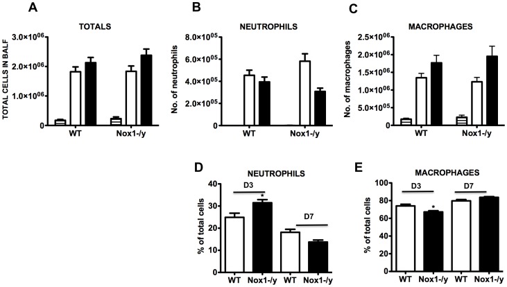
Figure 5. Effect of HkX-31 influenza A virus infection on BALF cellularity in WT and Nox1−/y mice.
Mice were treated with 1×10⁴ PFU of HkX-31 (H3N2) strain of influenza A virus and the number of (A) total cells, (B) neutrophils and (C) macrophages, and the percentage of (D) neutrophils and (E) macrophages counted in cytospin preparations of BALF 3 and 7 days post infection. In (A–C) Naïve (horizontal hash), HkX-31 (Day 3; open histogram) and HkX-31 (Day 7; filled histogram). Data are shown as mean ± SEM for 7–12 mice per group. *P<0.05 vs WT (ANOVA and Dunnett’s post hoc test).

