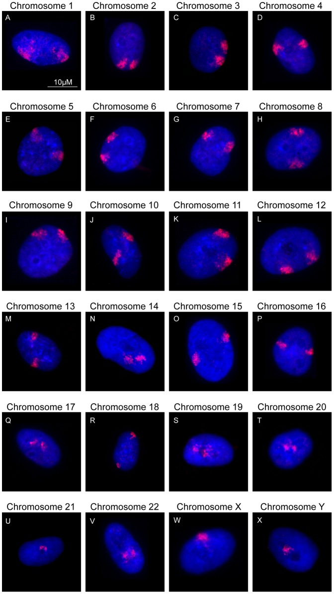
Figure 1. Positions of all chromosomes in normal proliferating human dermal fibroblasts:
Images displaying the spatial arrangement of each of the human chromosome territories (in red) in interphase nuclei (stained in blue) of fibroblasts. The numbers on the top of each nucleus indicates the chromosome to which a specific probe was hybridized to, as revealed by FISH. Scale bar = 10 µM.

